Free Online Productivity Tools
i2Speak
i2Symbol
i2OCR
iTex2Img
iWeb2Print
iWeb2Shot
i2Type
iPdf2Split
iPdf2Merge
i2Bopomofo
i2Arabic
i2Style
i2Image
i2PDF
iLatex2Rtf
Sci2ools
ICPR
2006
IEEE
2006
IEEE
Classification of Segmented Regions in Brightfield Microscope Images
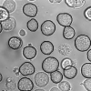
The subcellular localisation of proteins in living cells is an important step to determine their function. A common method is the evaluation of fluorescence images. The position of marked proteins, visible as bright spots, enables conclusions concerning their function. In order to determine the subcellular localisation, it is crucial to know the exact positions of the considered cells within an image. These are provided by the segmentation of a corresponding brightfield microscope image. As the resulting segments do not exclusively comprise cells, they have to be classified. Therefore, we propose an approach for the classification of the resulting segments in `cells' and `non-cells', which is an essential step of the automatic recognition of cells and thus of the automatic subcellular localisation of proteins in living cells.
Automatic Subcellular Localisation | Computer Vision | Corresponding Brightfield Microscope | ICPR 2006 | Marked Proteins |
Related Content
| Added | 09 Nov 2009 |
| Updated | 09 Nov 2009 |
| Type | Conference |
| Year | 2006 |
| Where | ICPR |
| Authors | Marko Tscherepanow, Frank Zöllner, Franz Kummert |
Comments (0)

